NEWSKU-36-MVZBC5KU-3XXS
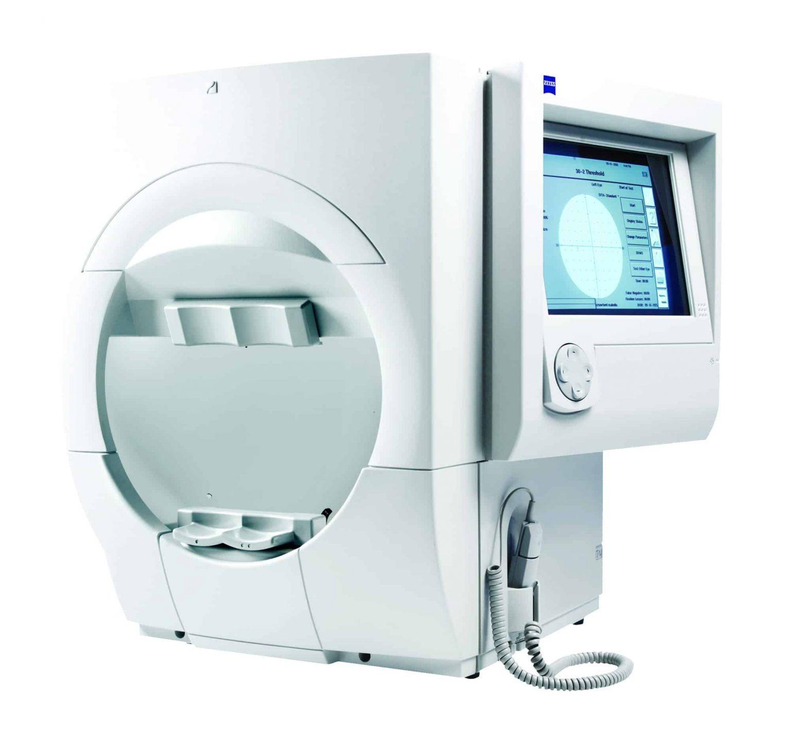
Computerized Visual Field Analyzer (CVF) – Automatic Ophthalmic Perimeter
Unit Price · Ex-Lagos
₦9,000,000
approx. $5,882.35 at today's rate
100 units available · MOQ 1 unit · Ships in 48hrs
× ₦9,000,000
Buyer protected Nationwide logistics Paystack verified
Fulfilled by Store
HH-00016
The Computerized Visual Field Analyzer (CVF) is an advanced ophthalmic diagnostic instrument designed to assess peripheral and central vision through automated visual field testing. It helps detect, evaluate, and monitor visual field defects associated with glaucoma, optic nerve disorders, retinal diseases, and neurological conditions.
Equipped with automated testing programmes, digital analysis software, and detailed visual field reporting capabilities, the equipment supports accurate eye examination and long-term patient monitoring. Suitable for ophthalmology clinics, optometry centres, eye hospitals, and diagnostic facilities.
Key Features & Specifications
- Equipment Type: Computerized Visual Field Analyzer
- Measurement Technology: Automated static perimetry
- Testing Method: Threshold and suprathreshold visual field testing
- Visual Field Range: Central 10°, 24° and 30° programmes; peripheral testing on supported models
- Test Patterns: 10-2, 24-2, 30-2 and other model-dependent patterns
- Stimulus Type: White-on-white light stimulus
- Stimulus Size: Goldmann stimulus sizes, depending on model
- Stimulus Duration: Typically 200 milliseconds
- Test Strategies: Full Threshold, Fast Threshold or manufacturer-specific automated strategies
- Fixation Monitoring: Automatic fixation monitoring on supported models
- Eye Tracking: Available on selected models
- Display: Digital LCD monitor or integrated touchscreen
- Patient Positioning: Adjustable chin rest and forehead support
- Testing Modes: Monocular testing; binocular programmes on supported models
- Glaucoma Analysis: Visual field defect assessment and progression monitoring
- Reliability Indicators: Fixation losses, false-positive and false-negative responses, where supported
- Reporting: Grey-scale maps, numerical sensitivity plots and statistical analysis
- Data Storage: Electronic patient database
- Printer: Integrated or external printer, depending on model
- Connectivity: USB, LAN or other available data interfaces
- Power Supply: Typically AC 100–240V, 50/60Hz
- Applications: Glaucoma assessment, visual field defect evaluation, optic nerve assessment and neurological visual field testing